
Tender, scaly, growing? Get it checked.
Squamous Cell Carcinoma (SCC) Treatment in Mount Gravatt, Brisbane
SCC is a skin cancer that can grow over weeks. Our specialist dermatologists remove most SCCs in our rooms, and treat biopsy-proven Bowen's disease (early SCC) with curettage and cautery or non-surgical laser-assisted PDT.
Specialist consultation $395. Medicare rebate applies with a valid GP referral. Surgery is quoted in writing before the procedure. Laser-assisted PDT is priced on assessment. · See fees
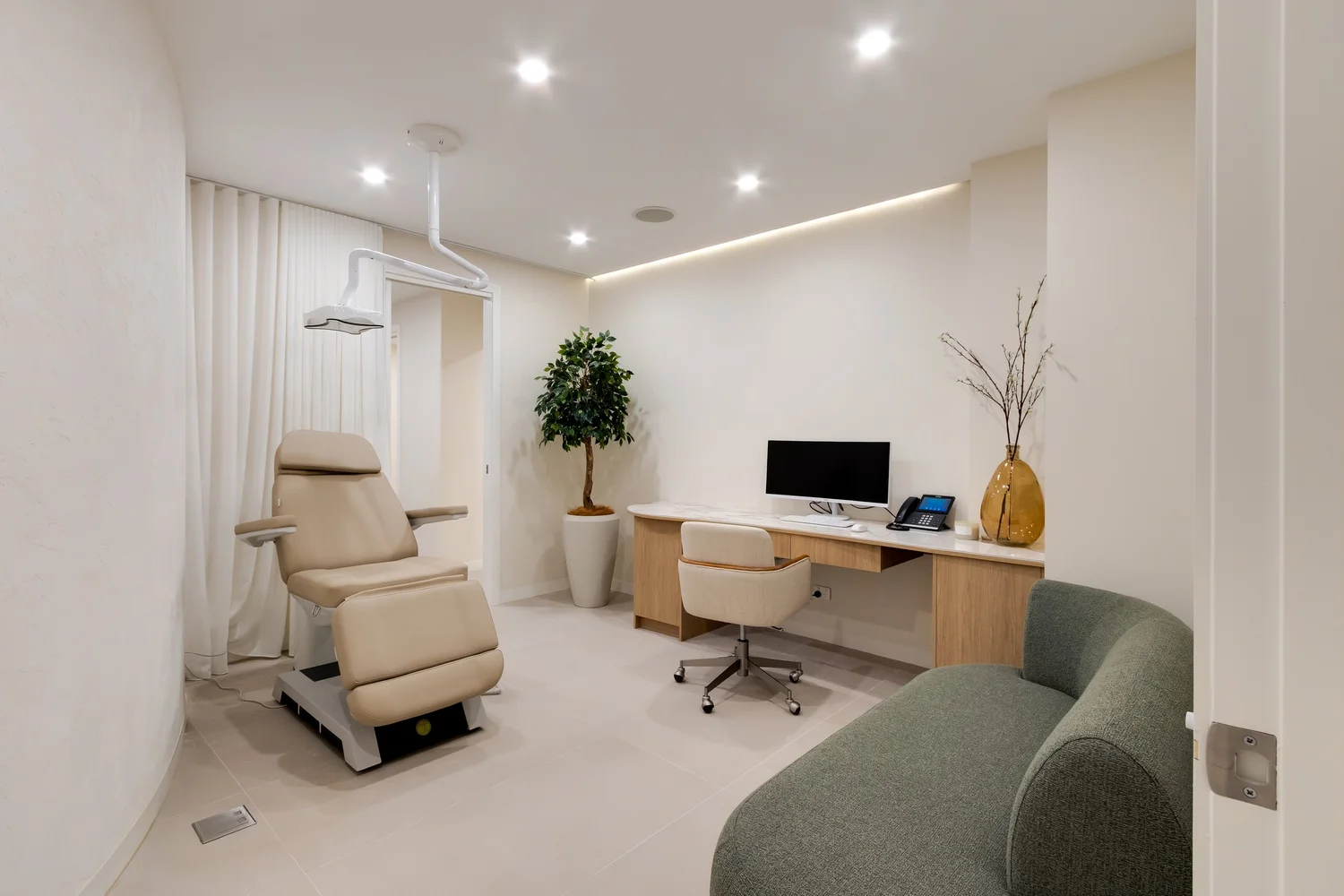
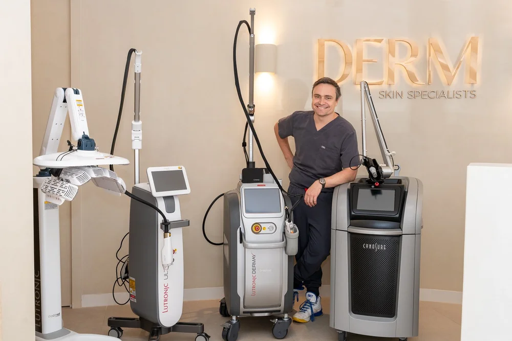
A dermatologist-led skin cancer clinic, founded by Dr Mitch Robinson, FACD, first author of Australia's evidence-based clinical practice guidelines for lentigo maligna.
Meet Dr MitchSCC Treatment at a Glance
What Is Squamous Cell Carcinoma (SCC), and How Common Is It in Australia?
The short answer: squamous cell carcinoma (SCC) is a skin cancer that starts in the flat (squamous) cells of the skin's top layer, not the pigment cells that cause melanoma. It's Australia's second most common skin cancer, after BCC. SCC usually grows on sun-damaged skin, often among rough sunspots, and can grow over weeks or months. Its earliest form, Bowen's disease, can often be treated without surgery.
SCC makes up about 30% of non-melanoma skin cancers (Cancer Council Australia), and about half a million Australians a year have a BCC or SCC surgically removed, with higher rates in Queensland (AIHW, 2026).
How it works
How an SCC is cut out
Every excision takes the cancer with a margin of normal skin, as an eye shape that closes flat.
- An invasive SCC ready to be removed. Your skin creases decide which way the cut will run.
- We mark a margin of normal-looking skin, usually 3 to 5 mm for an invasive SCC, and wider for larger or higher-risk SCCs.
- An eye shape is drawn around the margin, about three times longer than it is wide and lined up with your skin creases, so the wound closes flat.
- After numbing, the skin is cut out through its full thickness. The lab checks the SCC and confirms the edges are clear.
- The wound is closed in a straight line: dissolving stitches underneath, finer stitches on the surface. Stitches come out after about 5 to 7 days on the face and 10 to 14 on the body.
Simplified illustration, not to scale. Your margin and repair depend on the cancer and where it is.
Know the signs.
What Does SCC Look Like, and How Fast Does It Grow?
Cancer Council Australia lists the common signs of SCC as a thick scaly spot, a fast-growing pink lump, or a red, scaly or crusted spot. SCCs often feel tender, may bleed, and generally grow over weeks or months (Cancer Council Australia).
Clues that a spot could be an SCC:
- It hurts when you bump it or brush your hair
- A crust falls off and grows back thicker
- A scaly lump that wasn't there a couple of months ago
SCCs mostly appear on the head, neck, ears, lips, hands, forearms and lower legs.
SCC in olive, Asian and darker skin. SCC is less common in darker skin, and more often appears on skin that gets little sun, or in an old scar, burn or long-standing wound.
Bowen's Disease (SCC in Situ) vs Invasive SCC: What's the Difference?
Bowen's disease is SCC in situ: the cancer cells are confined to the top layer of skin. Pathology reports often call it intraepidermal carcinoma (IEC). It usually looks like a slowly growing, flat, red, scaly patch, often on the lower legs, and is easily mistaken for eczema or psoriasis.
Invasive SCC has grown into the deeper layer of skin (the dermis). It's usually raised, thicker and more often tender, and it's the form of SCC that can spread.
Why the difference matters:
- Bowen's disease can't spread while it stays in the top layer, so non-surgical treatment is an option.
- Left untreated, some Bowen's disease becomes invasive SCC.
- A thick, tender or fast-growing area in a Bowen's patch can mean it has become invasive, so we biopsy it.
How it works
Skin biopsy: shave or punch
A small sample, taken under local anaesthetic, tells the lab exactly what a spot is.
- A raised spot that needs a diagnosis, such as a possible BCC.
- A small injection of local anaesthetic numbs the skin. It stings for 10 to 20 seconds, then the area goes numb.
- A fine, flexible blade shaves off a thin disc of the spot through the top layers of skin.
- The sample goes to the lab, which confirms what the spot is, including its subtype. Results usually take about a week.
- The shallow wound heals like a graze, without stitches. If it’s a skin cancer, the result tells us how best to treat it.
- A spot that needs a diagnosis.
- Local anaesthetic numbs the area. The injection stings for 10 to 20 seconds, then the skin goes numb.
- A small round blade, like a tiny cookie cutter, is turned gently to take a core of skin a few millimetres wide.
- The core goes to the lab, which confirms what the spot is. Results usually take about a week.
- A stitch or two closes the small hole, and it heals as a small line.
Simplified illustration, not to scale.
No cut, no stitches.
Laser-Assisted PDT for Bowen's Disease: Non-Surgical Treatment in Our Clinic
For biopsy-proven Bowen's disease, laser-assisted photodynamic therapy (PDT) treats the patch without surgery. A dermatologist performs laser-assisted PDT for Bowen's disease in our Mount Gravatt clinic.
How it works, step by step:
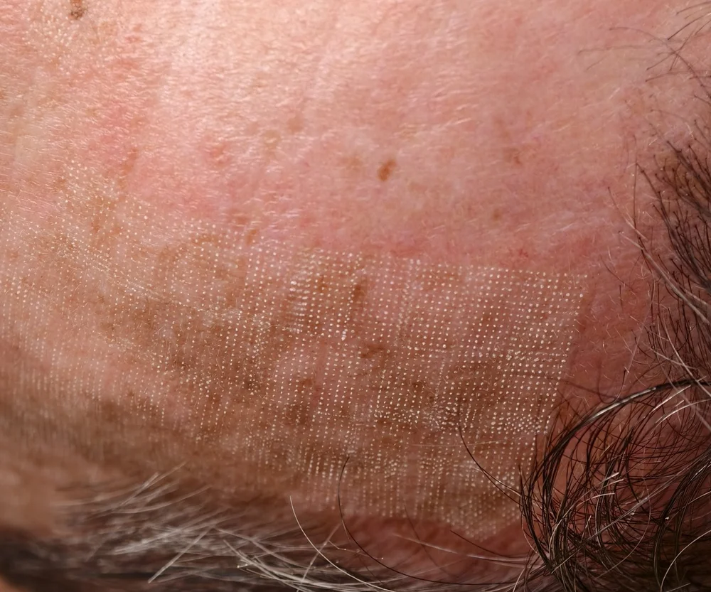
- Numbing and laser pass. We inject local anaesthetic into the patch. Then a quick pass of a fractional laser, either our CO2 laser or the LaseMD ULTRA, makes tiny channels in the top layer of skin over the Bowen's patch. This step is designed to help the light-sensitising cream penetrate to the abnormal squamous cells.
- Cream. The cream goes on the patch and stays covered for about an hour.
- Red light. About 30 minutes under red light from our Healite II LED panel activates the cream, which destroys the abnormal cells.
- Second session. Bowen's disease usually needs 2 sessions, 6 weeks apart.
- Follow-up. We check the patch has cleared, then keep watching it at your skin checks.
Afterwards: the patch is red and crusty for about a week, a little more than after standard PDT. Avoid strong light for about 48 hours after each session.
Who it suits:
- Biopsy-proven Bowen's disease (intraepidermal carcinoma)
- Large Bowen's patches, or patches in awkward spots
- Patches where you'd rather avoid a scar or a wound that heals slowly
Who it doesn't suit: invasive SCC, keratoacanthoma, and Bowen's patches that are thick, tender, fast-growing or have come back after treatment. These need surgery.
PDT leaves no tissue for the lab to check, so a small Bowen's patch in an easy spot is often simplest to cut out. See our PDT page.
Depth decides.
How Is Squamous Cell Carcinoma Treated? Options for Bowen's Disease and Invasive SCC
The options depend mostly on how deep the SCC goes.
Bowen's disease (SCC in situ)
- Laser-assisted PDT: no cut, usually 2 sessions 6 weeks apart, about a week of redness and crusting; needs follow-up
- Curettage and cautery: Dr Mitch routinely uses it for Bowen's disease away from the head and neck. The patch is scraped away and the base sealed with heat in one visit, with no stitches, and the scrapings go to the lab
- Surgical excision: often simplest for a small patch, and the lab confirms it has cleared
- Prescription creams: several weeks of home treatment with a red, crusty reaction; follow-up needed
- Freezing (cryotherapy): we also treat Bowen's disease by freezing it, with no cut and no stitches
How it works
Curette and cautery for Bowen’s disease
Scrape, seal, repeat. Quick, done under local anaesthetic, and no stitches.
- A patch of Bowen’s disease (an early SCC in the top layer of skin) away from the head and neck.
- Local anaesthetic numbs the area first.
- A sharp, spoon-shaped instrument called a curette scrapes out the soft cancer tissue. The scrapings go to the lab.
- Heat seals the base of the wound.
- Scraping and sealing are usually repeated two or three times in the one visit.
- There are no stitches. The wound heals on its own and usually leaves a small, flat, white scar.
Simplified illustration, not to scale. It suits selected small, superficial cancers.
Invasive SCC
- Surgical excision: the main treatment; Cancer Council's guidance is complete excision, with the lab confirming the SCC has been cleared
- Radiotherapy: when surgery isn't suitable, or after surgery for some high-risk SCCs
- Not suitable: PDT and creams, which don't reach deep enough for invasive SCC
Fast lump, central plug.
What Is a Keratoacanthoma, and Is It an SCC?
A keratoacanthoma is a dome-shaped lump with a crater or plug of hard scale (keratin) in the centre. It appears over a few weeks, often on the face, forearms or backs of the hands.
Some keratoacanthomas shrink on their own. But a keratoacanthoma can look very like an invasive SCC, to the eye and under the microscope, and the two can't always be told apart. So dermatologists usually treat a keratoacanthoma as an SCC and remove it. We don't use PDT for keratoacanthomas.
How Is SCC Diagnosed? Biopsy and What We Check First
At your consultation, your dermatologist will:
- Ask how fast the spot has grown, whether it's tender, and about past SCCs and your immune system.
- Examine it with a dermatoscope for the scale, keratin and blood vessel patterns of SCC and Bowen's disease.
- Feel the lymph nodes near an SCC on the head, neck, lip or ear.
- Check the rest of your skin, because SCCs grow among sunspots and other sun damage. See skin cancer and melanoma checks.
Biopsy. A small biopsy confirms an SCC and shows whether it's Bowen's disease or invasive. For invasive SCC, it also shows depth, differentiation (how abnormal the cells look) and any nerve involvement. Laser-assisted PDT needs a biopsy confirming Bowen's disease first.
Site and speed matter.
High-Risk SCC: Which Features Change the Plan, and When Do We Refer?
Cancer Council Australia's guidance lists features that raise the chance of an SCC coming back or spreading (Cancer Council quick reference guide):
- Site: the temple, ear or lip
- Size: wider than 20 mm
- Depth: thicker than 2 mm, or into the fat under the skin
- Nerves: SCC cells tracking along nerves (perineural invasion)
- Cell type: poorly differentiated SCC
- Immune system: long-term immune suppression, such as after an organ transplant or with chronic lymphocytic leukaemia
We also treat a recurrent SCC as high risk. A spreading SCC usually reaches nearby lymph nodes first; for head and neck SCCs, often in front of the ear (in the parotid gland) or in the neck.
When we refer:
- Head and neck or plastic surgeon: large or deep SCCs, enlarged lymph nodes, or complex sites around the eye, nose, lip or ear
- Radiation oncology: when surgery isn't suitable, or after surgery for nerve involvement or other high-risk features
- Medical oncology: the rare SCC that has spread, where immunotherapy may be considered
SCC Excision in Our Rooms: What's Different From Other Skin Cancer Surgery?
Local anaesthetic, stitches, healing, wound care, general risks and warning signs are covered on our skin cancer surgery page. What's specific to SCC:
- Margins: invasive SCC is usually removed with a 3–5 mm margin, which may be wider depending on size and high-risk features, with the lab checking the edges.
- Lips, ears and shins need planning: little spare skin often means a flap or graft, and complex lip and ear SCCs may be referred.
- Lower-leg wounds heal slowly, which is one reason Bowen's disease on the legs is usually treated with curettage and cautery or laser-assisted PDT rather than cut out.
- Don't wait for an event: SCCs can grow over weeks, so don't delay for a wedding or holiday; we'll plan around your dates where we can.
Higher risk, closer watch.
SCC in Organ Transplant Recipients and People With Weakened Immunity
After an organ transplant, or with a weakened immune system from a blood cancer or long-term immune-suppressing medicines, your risk of SCC is much higher. Cancer Council Australia rates chronic immune suppression, such as after a transplant or with chronic lymphocytic leukaemia, as very high risk (quick reference guide), and SCCs in people with weakened immune systems are more likely to spread (Cancer Council Australia).
What we do:
- Specialist skin checks at least yearly, often more frequently
- Biopsy or treat new scaly or tender spots early
- Treat Bowen's disease and sun-damaged skin early, to reduce new problem spots
Treat the soil, not just the weed.
Can Sunspots Turn Into SCC? Laser Solar Resurfacing for the Sun-Damaged Field
Occasionally, yes. Actinic keratoses (solar keratoses, often called sunspots) are precancerous, and a small number become SCC. The sun-damaged skin around them, called field change, is where many SCCs start, so after an SCC we look at the whole field.
Treating the field aims to reduce new sunspots and problem spots.
Laser solar resurfacing is our alternative to prescription field creams (weeks of red, crusty skin at home) and conventional PDT:
- LaseMD ULTRA, a 1927 nm fractional thulium laser, treats the top layer of the whole sun-damaged area. Dr Mitch uses it on its own to treat the field.
- About an hour of numbing cream, then about 15 minutes of laser
- Commonly 2 sessions about 6 weeks apart, each with about 4 to 5 days of redness and flaking
Laser solar resurfacing is a field treatment to reduce sunspots and new problem spots, not a treatment for an SCC itself. Any SCC or Bowen's patch is treated first, and thick sunspots are frozen or removed, before we treat the field.
More on sun damage treatment and LaseMD.
Know your nodes.
Will SCC Come Back? Recurrence, Lymph Node Checks and Follow-Up
An SCC can come back in or near its scar, or in the lymph nodes that drain it, and new SCCs tend to grow in the same sun-damaged skin. Cancer Council Australia's guidance is a skin check at least yearly after any BCC or SCC (Cancer Council quick reference guide). The high-risk features above, how many SCCs you've had and your immune system set how often we see you.
Between visits, check:
- The SCC scar or treated patch for a new lump, crust or sore
- Your lymph nodes: for head and neck SCCs, in front of and behind the ears, under the jaw and down the sides of the neck; the armpit for arm SCCs; the groin for leg SCCs
Call us about any new, firm lump that lasts more than two weeks.
How Much Does SCC Treatment Cost in Brisbane With Medicare?
- Specialist consultation: $395, with a Medicare rebate if you have a valid GP referral.
- SCC surgery: quoted in writing before the procedure. Medicare rebates apply to Medicare-listed skin cancer procedures.
- Laser-assisted PDT for Bowen's disease: price on assessment. It isn't covered by Medicare.
- Laser solar resurfacing: priced on assessment and quoted in writing.
- Pathology: billed separately by the lab.
See treatment fees and our skin cancer surgery page.
Who Treats SCC at DERM Skin Specialists?
SCC and Bowen's disease are diagnosed and treated by our specialist dermatologists (FACD): Dr Mitch Robinson, founder, Master of Skin Cancer (UQ), first author of Australia's lentigo maligna clinical practice guidelines, who leads our laser suite; Dr Emily Shao; and Dr Benny Yau. Dr Jimmy Wang (GP, FRACGP) does lower-priced GP-led skin checks and refers suspected SCCs to our dermatologists. We're in Mount Gravatt, a short drive from Greenslopes, Wishart and Eight Mile Plains.
Mount Gravatt, Brisbane
SCC Treatment in Mount Gravatt
DERM Skin Specialists is a dermatologist-led clinic at 2/1437 Logan Road, Mount Gravatt, with free onsite parking and wheelchair access. We're about 15 minutes from the Brisbane CBD and close to Holland Park, Carindale, Upper Mount Gravatt, Mansfield and Sunnybank. Patients are typically seen within 2 weeks.
Your questions, answered
SCC and Bowen's Disease FAQs
Is SCC more serious than a BCC?
Usually, yes. An SCC can grow over weeks and is more likely to spread, while most BCCs grow slowly and rarely spread. Both are usually cured when treated early.
Can an SCC go away on its own?
No. Some keratoacanthomas shrink, but because they can't always be told apart from SCC, we remove them.
Is Bowen's disease cancer?
Yes, an early one. Bowen's disease is SCC that hasn't grown below the top layer of skin.
Is intraepidermal carcinoma the same as Bowen's disease?
Yes. Intraepidermal carcinoma (IEC), SCC in situ and Bowen's disease are three names for SCC confined to the top layer of skin.
Why do you need a biopsy before PDT for Bowen's disease?
To confirm it's Bowen's disease, not invasive SCC or another skin cancer. PDT can't reach an invasive SCC, so it could leave cancer behind.
Can laser-assisted PDT treat an invasive SCC?
No. Invasive SCC has grown deeper than PDT can reach. It needs surgery, or radiotherapy when surgery isn't suitable.
Does laser-assisted PDT for Bowen's disease hurt?
We numb the patch first with local anaesthetic injected into it, before the laser step. You may still feel some warmth or stinging while the red light is on.
Can Bowen's disease be scraped off instead of cut out?
Often, yes. Away from the head and neck, Dr Mitch routinely treats Bowen's disease with curettage and cautery: the patch is scraped away and the base sealed with heat in one visit, with no stitches, and the scrapings go to the lab.
Can Bowen's disease come back after PDT?
It can, so we recheck the treated patch. A Bowen's patch that comes back usually needs surgery, so the lab can confirm it's gone.
What does "perineural invasion" mean on an SCC pathology report?
SCC cells were seen tracking along a small nerve. This raises the chance of the SCC coming back, so we may discuss radiotherapy and closer follow-up.
Should I stop my transplant medicines because of SCC?
No. Never stop or change anti-rejection medicines yourself; that's your transplant team's decision.
Is a cutaneous horn an SCC?
Sometimes. A cutaneous horn is a hard spike of keratin. Its base can be a harmless growth, a sunspot, Bowen's disease or an SCC, so we remove it for the lab.
Does daily sunscreen lower SCC risk?
Yes. Daily sunscreen on sun-exposed skin, with a hat, shade and covering clothing, helps lower your risk of new SCCs. Put it on every morning, not just on beach days.
Can a vitamin B3 supplement lower my SCC risk?
It can help some people. In people with normal immune systems who have had several non-melanoma skin cancers, a vitamin B3 supplement can lower the number of new ones, including SCCs, while they keep taking it. It hasn't been shown to help organ transplant recipients. Ask us first: the form and dose matter.
Worried a Tender, Scaly Spot Could Be an SCC?
SCCs can grow over weeks, so a sore that won't heal is worth checking sooner rather than later. Book with our specialist dermatologists in Mount Gravatt, including to see if laser-assisted PDT suits your Bowen's disease.
Clinical content overseen by Dr Mitch Robinson, FACD (AHPRA MED0001946771). Updated October 2026. General information only, not medical advice. All procedures carry risks; suitability, recovery and results vary between individuals.
Sources: Cancer Council keratinocyte cancer guidelines · Cancer Council: About skin cancer · Cancer Council GP quick reference guide · AIHW 2026