
Australia's most common skin cancer
Basal Cell Carcinoma (BCC) Treatment in Mount Gravatt, Brisbane
Specialist dermatologists diagnose and treat BCC in our Mount Gravatt clinic, with surgery, curettage, or non-surgical laser-assisted PDT for superficial and selected nodular BCCs.
Specialist consultation $395. Medicare rebate applies with a valid GP referral. Surgery fees are quoted in writing before treatment; laser-assisted PDT is priced on assessment. · See fees
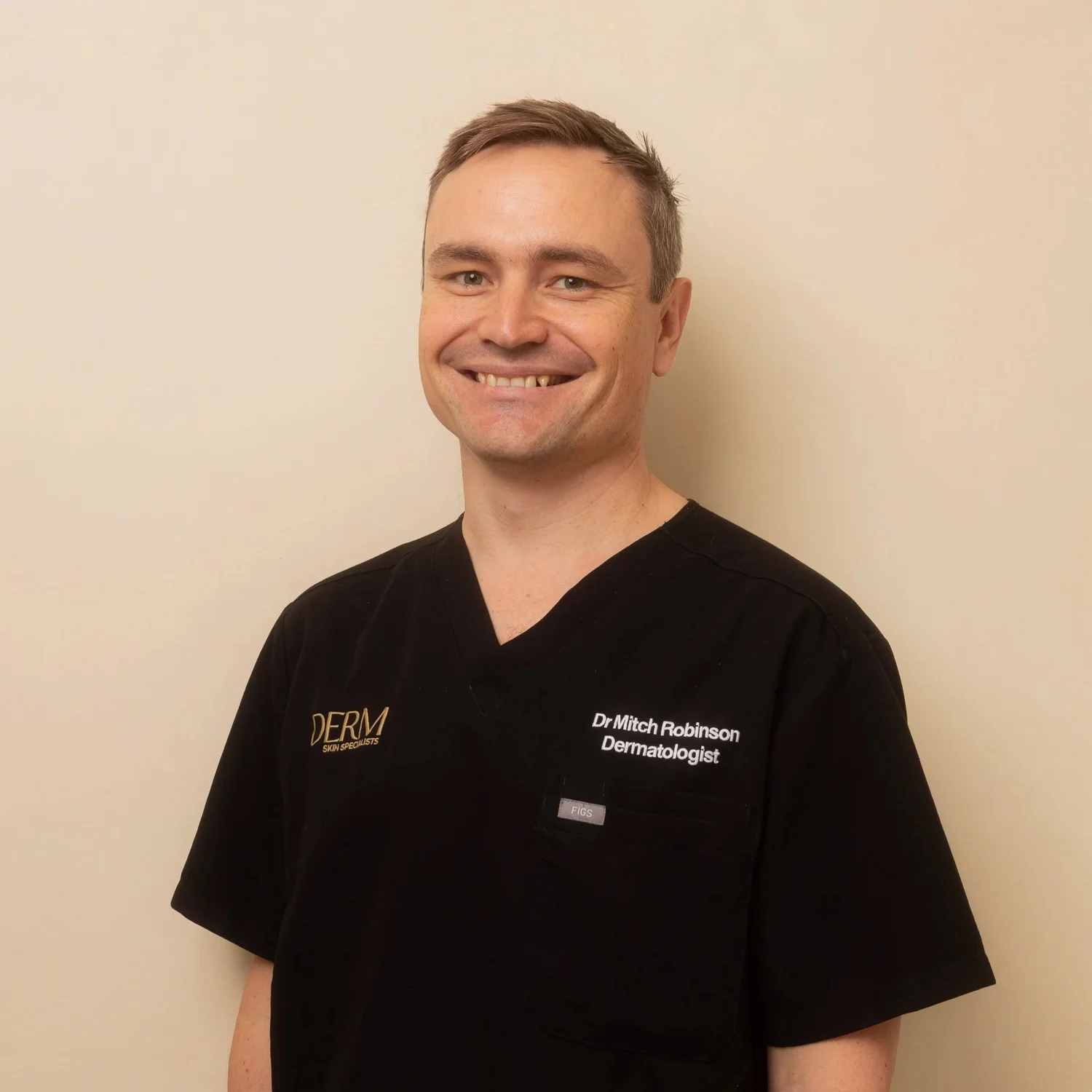
A dermatologist-led skin cancer clinic, founded by Dr Mitch Robinson, FACD, first author of Australia's evidence-based clinical practice guidelines for lentigo maligna.
Meet Dr MitchBasal Cell Carcinoma at a Glance
Most common. Very treatable.
What Is a Basal Cell Carcinoma (BCC), and Is It Dangerous?
The short answer: a BCC is a skin cancer that starts in the basal cells, at the bottom of the skin's outer layer. Cancer Council Australia says it makes up about 2 out of 3 skin cancers, grows slowly over months or years, and very rarely spreads. Most BCCs are cured when they're treated properly.
The danger is local. Left alone, a BCC keeps growing outwards and downwards and may invade nerves and tissue, making treatment harder. On the nose or eyelid, a few extra millimetres can mean a bigger repair.
How it works
How a BCC is cut out
Every excision takes the cancer with a margin of normal skin, as an eye shape that closes flat.
- A BCC ready to be removed. The natural creases of your skin decide which way the cut will run.
- We mark a margin of normal-looking skin around the BCC, usually a few millimetres, so the whole cancer comes out.
- The cut is drawn as a narrow eye shape about three times longer than it is wide, lined up with your skin creases, so it closes flat.
- After numbing, the eye shape is removed through the full thickness of the skin and sent to the lab, which checks the edges are clear.
- Dissolving stitches underneath take the tension, and finer stitches close the surface in a straight line. They come out after about 5 to 7 days on the face, 10 to 14 on the body.
Simplified illustration, not to scale. Your margin and repair depend on the cancer and where it is.
So it isn't an emergency, but don't leave it for months. Australia's optimal care pathway for keratinocyte cancer suggests specialist referral within 8 weeks for a presumed BCC. A thin, superficial BCC can often be treated without surgery.
Pearly, pink or scar-like.
What Does a BCC Look Like?
BCCs usually appear on sun-exposed skin: the face, nose, ears, neck, shoulders and upper back. The four main subtypes look different.
- Nodular BCC, the most common type: a pink or pearly, shiny lump with fine blood vessels on the surface. It may break down into a crusted sore.
- Superficial BCC: a flat, pink or red scaly patch, often on the chest, back, arms or legs, and easily mistaken for eczema.
- Infiltrative or morphoeic BCC: a pale, firm, shiny area that looks like a scar. It grows in thin strands under the skin, so it's often bigger than it looks.
- Pigmented BCC: contains brown, blue-grey or black colour, so it can look like a mole or even a melanoma.
A typical BCC story: a spot bleeds when you dry your face, scabs over, seems to heal, then breaks down again weeks later. That's worth a skin cancer check.
Biopsy first, then the plan.
BCC Biopsy First: Why the Subtype Decides Your Treatment
Dermoscopy, a handheld magnifying light, often shows the branching vessels and pearly edge of a BCC. The lab confirms it: a small punch or shave biopsy under local anaesthetic shows it's a BCC, and which subtype.
How it works
Skin biopsy: shave or punch
A small sample, taken under local anaesthetic, tells the lab exactly what a spot is.
- A raised spot that needs a diagnosis, such as a possible BCC.
- A small injection of local anaesthetic numbs the skin. It stings for 10 to 20 seconds, then the area goes numb.
- A fine, flexible blade shaves off a thin disc of the spot through the top layers of skin.
- The sample goes to the lab, which confirms what the spot is, including its subtype. Results usually take about a week.
- The shallow wound heals like a graze, without stitches. If it’s a skin cancer, the result tells us how best to treat it.
- A spot that needs a diagnosis.
- Local anaesthetic numbs the area. The injection stings for 10 to 20 seconds, then the skin goes numb.
- A small round blade, like a tiny cookie cutter, is turned gently to take a core of skin a few millimetres wide.
- The core goes to the lab, which confirms what the spot is. Results usually take about a week.
- A stitch or two closes the small hole, and it heals as a small line.
Simplified illustration, not to scale.
- Superficial BCC sits in the outer layers of skin, so surgery, laser-assisted PDT or curettage may suit, and occasionally a prescription cream.
- Nodular BCC goes deeper. It's usually cut out, or scraped out if it's small and on the body, and selected nodular BCCs can suit laser-assisted PDT.
- Infiltrative or morphoeic BCC spreads beyond its visible edge, so it usually needs a wider excision or Mohs surgery.
- Recurrent BCC, one that has come back after earlier treatment, usually needs surgery.
PDT and creams leave nothing for the lab to check, so a biopsy confirms the BCC's subtype before we use them.
Laser, cream, then light.
BCC Without Surgery: How Laser-Assisted PDT Works
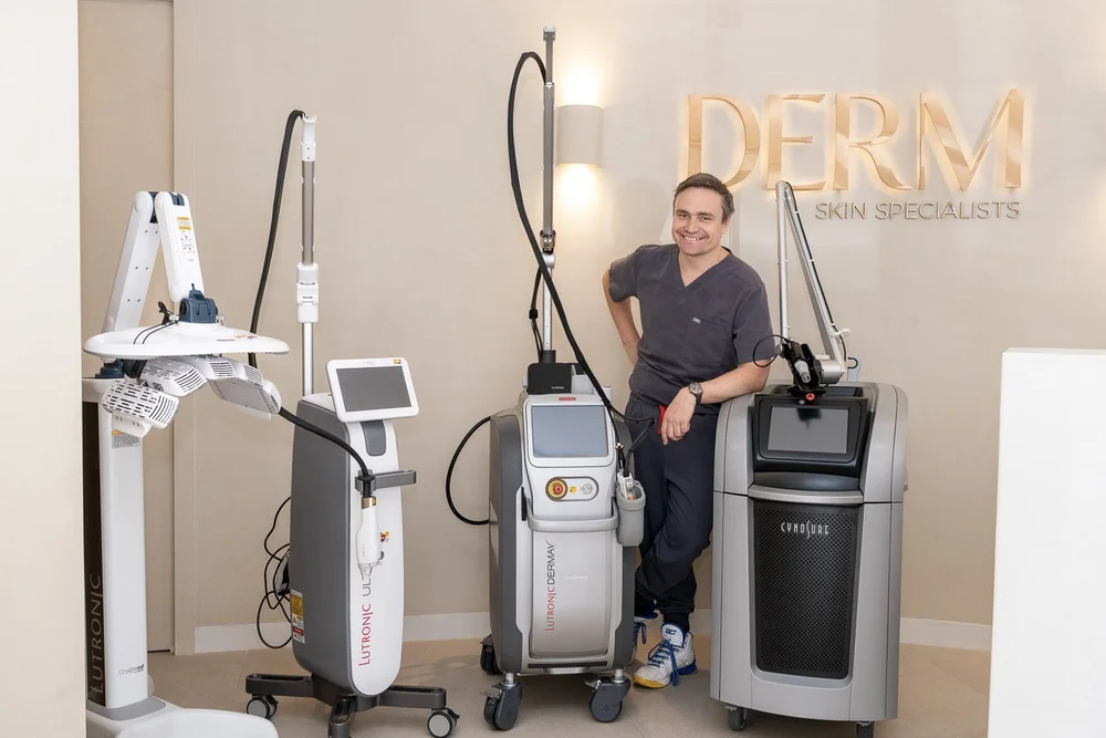
For a biopsy-proven superficial BCC, and selected nodular BCCs, laser-assisted photodynamic therapy (PDT) treats the cancer without surgery. A dermatologist performs laser-assisted PDT in our Mount Gravatt clinic, and Dr Mitch uses this approach for many skin cancer cases.
How it works
Laser-assisted PDT vs conventional PDT
Same cream, same red light. The difference is how far the cream gets in.
Laser-assisted PDT
- The area is cleaned, any scale is gently removed and the spot is numbed. A quick pass of a fractional laser, either our CO2 laser or the LaseMD ULTRA, then makes tiny channels in the top layer of skin.
- The light-sensitising cream goes on under a light-blocking dressing for about an hour. The channels let more of it reach the abnormal cells, which take up more cream than normal skin.
- Red light from our Healite II LED panel shines on the area for about 30 minutes. It switches the cream on, and the abnormal cells break down over the following days.
- Red and crusty for about a week, with a little more redness and flaking than conventional PDT. Stay out of strong light for about 48 hours. Skin cancers usually need 2 sessions, 6 weeks apart.
Conventional PDT
- The area is cleaned and any scale is gently removed, so the cream can soak in. There is no laser step.
- The same cream goes on under a light-blocking dressing for about an hour. It soaks in through the skin’s surface, so it mainly reaches abnormal cells near the top.
- The same red light, for about 30 minutes. Cells that took up the cream break down. Cells the cream barely reached are less affected.
- Red and crusty for about a week. Stay out of strong light for about 48 hours. Usually 1 session for sun damage, 2 for skin cancers. In thicker spots, deeper cells can remain, so we review you and re-treat or use another option.
Simplified illustration, not to scale. Suitability and results vary between people.
How it works, step by step
- Numbing and laser pass. We inject local anaesthetic directly into the BCC. Then a quick pass of a fractional laser, either our CO2 laser or the LaseMD ULTRA, makes tiny channels in the outer layer of skin.
- Light-sensitising cream. The cream goes on and is covered for about an hour. The channels are designed to help more of it reach the abnormal cells.
- Red light. Our Healite II LED panel shines red light on the area for about 30 minutes. The light activates the cream, and the BCC cells break down over the following days.
- Second session. A BCC usually needs 2 sessions, 6 weeks apart, then close follow-up.
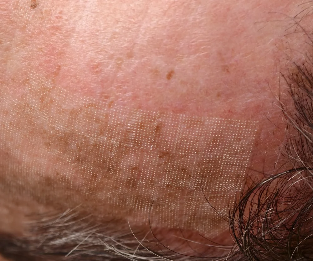
Who it suits: thin, low-risk superficial BCCs and selected nodular BCCs, especially where a scar or a slow-healing wound is a real concern. Suitability depends more on the BCC's type and depth than on where it is, and we decide case by case.
Who it doesn't suit: infiltrative or morphoeic BCCs, BCCs that have come back, and poorly defined BCCs. These usually need surgery. For a nodular BCC, we'll tell you whether yours is one that suits laser-assisted PDT.
PDT and surgery. Surgery has the highest cure rate for BCC, and the lab confirms the whole cancer is gone. PDT leaves no specimen, so follow-up visits do that job.
Published results. In a published UK series of 177 biopsy-proven BCCs, 97.1% were still clear an average of 32 months after CO2 laser-assisted PDT (Annals of Plastic Surgery, 2014), and the same unit reported 93.6% still clear at 5 years (Annals of the Royal College of Surgeons of England, 2021).
Recovery. Expect about a week of redness and crusting, a little more than after standard PDT. Avoid strong light, including sunlight, for about 48 hours. Colour change in the treated skin is possible. BCCs grow slowly, so treatment can often be timed around a wedding or holiday. Read more on our PDT page.
BCC Treatment Options Compared: Surgery, Laser-Assisted PDT, Curette or Cream
Surgery (excision)
- Suits: every BCC subtype and most sites, including the face
- Clearance: the highest cure rate, and the lab checks the whole BCC
- Healing: stitches, then a fine line scar
- Visits: one procedure in our rooms
Laser-assisted PDT
- Suits: thin, low-risk superficial BCCs and selected nodular BCCs
- Clearance: lower than surgery, with no lab check
- Healing: about a week of redness and crusting, usually little or no scarring
- Visits: usually 2 sessions, 6 weeks apart, then follow-up
Curettage and cautery
- Suits: small, well-defined nodular or superficial BCCs on the body
- Clearance: the scrapings go to the lab, but the edges can't be checked as they are after excision
- Healing: an open wound for about 2 to 4 weeks, then a flat, round, paler scar
- Visits: one
Prescription cream
- Suits: biopsy-proven superficial BCCs, not deeper types
- Clearance: lower than surgery, with no lab check
- Healing: several weeks of red, sore, crusty skin at home, often with little visible mark
- Visits: applied at home, then a review
BCC Excision: Why Surgery Is Usually First Choice
Australia's optimal care pathway for keratinocyte cancer recommends complete excision for most lesions. For a BCC, that means cutting it out with a margin of normal-looking skin and sending it to the lab.
- It's the main treatment for nodular, infiltrative and recurrent BCCs, and for most BCCs on the face.
- Pathology confirms the subtype and whether the edges are clear. If they aren't, we'll discuss the next step.
- Most BCC removal is done in our rooms under local anaesthetic, sometimes with a flap or graft on the nose or face.
Numbing, stitches, healing, risks and warning signs are the same as for any skin cancer operation: see our skin cancer surgery page.
Curettage and Cautery for BCC: Who Suits It?
Curettage and cautery means scraping out the BCC with a sharp, spoon-shaped instrument, then sealing the base with heat, usually two or three times in one visit. Dr Mitch uses it for suitable BCCs, and Cancer Council's guide for GPs lists it as an option for superficial and nodular BCCs under 2 cm.
How it works
Curette and cautery for a BCC
Scrape, seal, repeat. Quick, done under local anaesthetic, and no stitches.
- A small, well-defined BCC on the trunk, an arm or a leg.
- Local anaesthetic numbs the area first.
- A sharp, spoon-shaped instrument called a curette scrapes out the soft cancer tissue. The scrapings go to the lab.
- Heat seals the base of the wound.
- Scraping and sealing are usually repeated two or three times in the one visit.
- There are no stitches. The wound heals on its own and usually leaves a small, flat, white scar.
Simplified illustration, not to scale. It suits selected small, superficial cancers.
- It can suit: small, well-defined BCCs on the trunk, arms or legs, and people with several BCCs.
- It doesn't suit: infiltrative or morphoeic BCCs, BCCs that have come back, or most BCCs on the central face.
It's quick, with no stitches.
Small spot, tricky site.
BCC on the Nose and Face: What Changes?
The nose is one of the most common places for a BCC. There it often looks like a small, shiny, pearly bump, a spot that bleeds when you blow your nose, or a sore that keeps scabbing.
The skin there is thin and tight over cartilage. Cancer Council's guide for GPs lists the lips, nose, ears, eyelids and scalp as anatomically significant sites, and suggests considering Mohs surgery for morphoeic BCCs, particularly on the nose or the crease beside it.
For a BCC on the nose or face, we confirm the subtype with a biopsy in most cases, plan the margin and repair together so the nose keeps its shape, and refer for Mohs surgery when the subtype or site calls for it. Whether laser-assisted PDT suits a BCC on the face depends on its type and depth, so we talk through surgical and non-surgical options together.
When Is a BCC Referred for Mohs Surgery or Radiotherapy?
Mohs surgery removes the BCC in thin layers, checking the whole edge under the microscope the same day, to save as much normal skin as possible. We refer for Mohs when it adds real value: infiltrative or poorly defined BCCs on the face, recurrent BCCs, and BCCs on the eyelid, nose tip, lip or ear.
Radiotherapy for a BCC is given by a radiation oncologist over a series of visits. Australia's optimal care pathway recommends it for people who decline or can't have surgery, and after surgery in some higher-risk situations. Treated skin can change in texture and colour over the years.
Rarely, a BCC becomes very large or spreads, and specialist cancer teams may use targeted tablets or immunotherapy.
For the skin around the scar.
After a BCC, Can Laser Solar Resurfacing Reduce New Sunspots?
The years of sun behind your BCC often leave rough, scaly sunspots (actinic keratoses) nearby, in sun-damaged skin called field change. A small number of sunspots become squamous cell carcinomas.
Laser solar resurfacing is our in-clinic alternative to prescription field creams and conventional PDT for that field. It's a field treatment, not a treatment for the BCC itself: we treat your BCC first, then the field.
- What it is: the LaseMD ULTRA, a 1927 nm fractional thulium laser, treats the whole area.
- On the day: about an hour of numbing cream, then about 15 minutes of laser.
- Sessions: commonly 2 sessions, about 6 weeks apart.
- Recovery: about 4 to 5 days of redness and flaking.
- The aim: fewer new sunspots, alongside regular skin checks.
One BCC raises the odds of another.
Can a BCC Come Back After Treatment?
Sometimes, in two ways:
- The same BCC comes back in or near the treated spot. This is less likely after a clear excision, and more likely after PDT, creams or curettage, and with infiltrative BCCs.
- A new BCC appears somewhere else. Cancer Council Australia notes that having had a BCC increases the risk of another.
So Australia's optimal care pathway advises at least a yearly skin check after a skin cancer, and every 6 to 12 months if you're at higher risk.
Between visits, watch the treated spot: a pearly lump, scab or sore returning there is the classic sign of a BCC coming back. Call 07 3487 1414 if you notice one.
BCC in Younger People and in Olive, Asian or Darker Skin
BCC is more common after 40, but Cancer Council Queensland notes it can develop in young people. In your 20s or 30s, a scaly pink patch on the back or chest is easy to blame on eczema, so one that keeps flaring in the same place is worth checking. A few rare inherited conditions cause many BCCs from a young age.
In olive, Asian and darker skin, BCC is less common and more often pigmented, so it can be mistaken for a mole. On dermoscopy, a pearly edge and visible blood vessels help tell them apart. Darker skin is also more prone to colour change or thickened scars, which we factor into the choice of BCC treatment.
Quoted before you start.
What Does BCC Treatment Cost, Including Laser-Assisted PDT?
- Specialist consultation: $395, with a Medicare rebate if you have a valid GP referral.
- Excision or curettage of a BCC: Medicare rebates apply to Medicare-listed skin cancer procedures, and your fee is quoted in writing before the procedure.
- Laser-assisted PDT: price on assessment. It isn't covered by Medicare.
- Pathology on your biopsy or excision: billed separately by the lab.
See treatment fees, and the Medicare section of our skin cancer surgery page.
Dermatologist-led, in our rooms.
Who Diagnoses and Treats Your BCC in Mount Gravatt?
Your BCC is diagnosed and treated by our specialist dermatologists (FACD): Dr Mitch Robinson, Dr Emily Shao and Dr Benny Yau. A dermatologist performs laser-assisted PDT in our Mount Gravatt clinic. Dr Mitch holds a Master of Skin Cancer (UQ), leads our laser suite and teaches laser to dermatology registrars.
Dr Jimmy Wang (GP, FRACGP) offers lower-priced GP-led skin checks and refers suspected BCCs to our dermatologists. We see patients from Holland Park, Carindale, Sunnybank, Mansfield and across Brisbane's southside, with free onsite parking.
Mount Gravatt, Brisbane
BCC Treatment in Mount Gravatt
DERM Skin Specialists is a dermatologist-led clinic at 2/1437 Logan Road, Mount Gravatt, with free onsite parking and wheelchair access. We're about 15 minutes from the Brisbane CBD and close to Holland Park, Carindale, Upper Mount Gravatt, Mansfield and Sunnybank. Patients are typically seen within 2 weeks.
Your questions, answered
BCC Frequently Asked Questions
My spot scabbed over and healed. Could it still be a BCC?
Yes. A BCC can seem to heal, then scab or bleed again weeks later. It doesn't go away on its own, and keeps slowly growing until it's treated.
Can a BCC turn into melanoma?
No. A BCC starts in basal cells and melanoma in pigment cells. But a BCC shows enough sun damage to raise your risk of other skin cancers, so a full skin check matters.
Is there a cream that treats BCC?
For a biopsy-proven superficial BCC, a prescription cream used at home for several weeks is one option, with red, crusty skin, no lab check and close follow-up. Creams don't suit deeper BCCs, and over-the-counter creams don't treat BCC.
Is laser-assisted PDT the same as having a BCC lasered off?
No. The laser doesn't remove the BCC. It makes tiny channels so more light-sensitising cream reaches the abnormal cells, then red light activates the cream.
How many laser-assisted PDT sessions does a BCC need?
Usually 2, 6 weeks apart, then close follow-up to check the BCC has cleared.
My BCC is nodular. Is PDT still an option?
Sometimes. Laser-assisted PDT can suit selected nodular BCCs, and after the biopsy we'll tell you whether yours is one. Other nodular BCCs are usually cut out or, if small and on the body, treated with curettage and cautery.
Will laser-assisted PDT on my BCC sting?
We numb the BCC first with local anaesthetic injected directly into it, before the laser step. You may still feel some warmth or stinging while the red light is on.
What if my BCC returns after PDT?
A BCC that returns is no longer suited to PDT. It's usually removed surgically, and a recurrent BCC on the face may be referred for Mohs surgery.
Can laser-assisted PDT treat a BCC on my nose?
It depends on the BCC more than where it is. If the biopsy shows a thin superficial BCC or a suitable nodular BCC, laser-assisted PDT may be an option. Infiltrative BCCs usually need surgery wherever they are.
Will laser solar resurfacing stop me getting another BCC?
No treatment can promise that. Laser solar resurfacing treats sunspots and sun-damaged skin to reduce new sunspots. It doesn't treat a BCC or replace regular skin checks.
Can my GP treat my BCC instead?
Often, yes. Many GPs treat simple BCCs. A dermatologist is worth considering for a facial, large, recurrent or infiltrative BCC, many BCCs, or to discuss laser-assisted PDT.
My pathology report says superficial BCC. What does that mean?
It means the BCC is growing as a thin, flat layer near the skin surface, not as a deeper lump. It's the subtype that most often suits laser-assisted PDT. "Infiltrative" or "mixed" on the report usually means surgery, and for "nodular" we'll talk through whether laser-assisted PDT suits.
Is laser-assisted PDT for a BCC covered by Medicare?
No. Laser-assisted PDT isn't covered by Medicare. It's priced on assessment, after your consultation.
Worried a Spot Might Be a BCC?
A pearly bump or a sore that keeps scabbing is worth checking. Ask whether your BCC suits surgery or laser-assisted PDT. Patients are typically seen within 2 weeks.
Clinical content overseen by Dr Mitch Robinson, FACD (AHPRA MED0001946771). Updated October 2026. General information only, not medical advice. All procedures carry risks; suitability, recovery and results vary between individuals.
Sources: Cancer Council: About skin cancer · Cancer Council Queensland · Optimal care pathway: keratinocyte cancer · Cancer Council GP quick reference guide · Shokrollahi et al. Ann Plast Surg 2014 · Azzopardi et al. Ann R Coll Surg Engl 2021