
Found early. Treated properly.
Melanoma Diagnosis and Treatment in Mount Gravatt, Brisbane
Specialist dermatologists who diagnose melanoma with dermoscopy, confirm it with the right biopsy, and treat it following the Australian melanoma guidelines.
Specialist consultation $395. Medicare rebate applies with a valid GP referral. Surgery fees quoted in writing before the procedure. · See fees
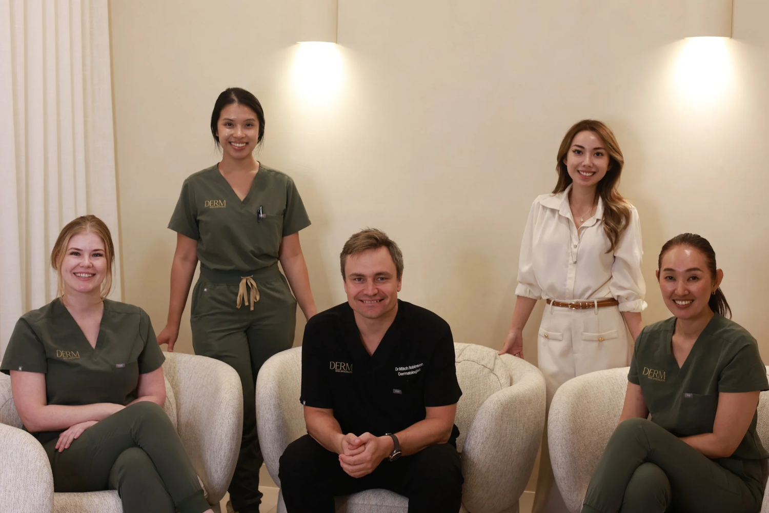
A dermatologist-led skin cancer clinic, founded by Dr Mitch Robinson, FACD, first author of Australia's evidence-based clinical practice guidelines for lentigo maligna.
Meet Dr MitchMelanoma Diagnosis and Treatment at a Glance
Know what you're dealing with.
What Is Melanoma, and How Common Is It in Queensland?
The short answer: melanoma is a cancer of melanocytes, the cells that make skin pigment. It's less common than BCC or SCC, but it's the skin cancer most likely to spread. Found early, it's usually cured with a simple operation.
How it works
Melanoma: excision biopsy, then wide local excision
The first excision confirms the melanoma and its thickness. The second takes the margin that thickness calls for.
- A raised spot that might be melanoma is removed whole, not sampled, so the pathologist can see all of it.
- An excision biopsy takes the spot with a 2 mm margin of normal skin, drawn as a small eye shape.
- The spot goes to the lab and the wound is closed with a few stitches. It’s done in our rooms under local anaesthetic and usually takes 20 to 30 minutes.
- The pathologist measures how deep the melanoma goes, its Breslow thickness. That sets the next margin. Results usually take about a week.
- The wide local excision margin is measured from the biopsy scar: 5 to 10 mm for melanoma in situ, 1 cm up to 1 mm thick, 1 to 2 cm from 1 to 4 mm, and 2 cm over 4 mm.
- The wider eye shape is removed for the lab, and the wound is closed in a line. On tight skin, such as the face, shins or scalp, it may need a flap or graft.
Simplified illustration, not to scale. Your margin and repair depend on the cancer and where it is.
Australia has one of the highest melanoma rates in the world. The Australian Institute of Health and Welfare (AIHW) estimates around 17,400 new melanoma cases in 2025. Queensland has historically had higher rates than the national average, with the highest state rate in 2021 at 79 cases per 100,000 people (AIHW).
It isn't only an older person's cancer. AIHW estimates melanoma is the second most common cancer in Australians under 40. Melanoma Institute Australia reports that about 95% of melanomas are caused by UV exposure, and that about a third grow from an existing mole, while the rest appear as new spots (Melanoma Institute Australia).
Melanoma can affect anyone. Fair skin, many moles, a history of sunburn, a past melanoma or a family history all raise the risk. Olive, Asian and darker skin has lower risk, but not zero.
Not every melanoma is dark.
What Does Early Melanoma Look Like?
Most people know to watch for a dark, irregular, changing mole. Our skin checks page covers the ABCDE signs and dermoscopy basics. Here's what's worth knowing beyond that.
Early melanoma is often flat. It may be a mole that looks different to your other moles (the "ugly duckling"), a spot that's slowly spreading, or a mole with a new colour such as blue-grey, red or white.
Some melanomas don't follow the rules at all. A summary in the Medical Journal of Australia, part of the Australian melanoma guideline series, notes that up to 20% of melanomas are only partly pigmented, and some have no pigment at all (Australian melanoma guidelines, MJA 2017). These can look like a pink pimple, a scar or a small pink lump.
The single most useful sign is change. A spot that is new, growing or changing over more than a month deserves a specialist look.
Five shapes, one disease.
What Are the Different Types of Melanoma?
Melanoma is grouped into types based on how it grows. The type changes what it looks like, where it turns up, and how fast it can thicken.
Superficial Spreading Melanoma
The most common type. It grows outwards along the skin before it grows down, often over months to years. It usually looks like a flat, irregular brown or black patch with uneven colour, often on the back in men and the legs in women.
Nodular Melanoma and the EFG Rule
Nodular melanoma grows downwards quickly and may skip the flat phase. It's often a firm, raised, dome-shaped lump that can be black, red, pink or skin-coloured.
Because nodular melanoma often fails the ABCDE checklist, Australian authors use EFG: Elevated, Firm and Growing. The melanoma guideline summary advises that any lump that is elevated, firm and growing for more than a month should be checked promptly and is usually excised rather than watched (Australian melanoma guidelines, MJA 2017).
Lentigo Maligna Melanoma
This grows on sun-damaged skin, usually the face, ears and scalp of older adults. It starts as lentigo maligna, a slowly enlarging flat brown patch that's easily mistaken for a sun spot. When it invades deeper, it's called lentigo maligna melanoma. Dr Mitch was first author of the Australian clinical practice guidelines for lentigo maligna. See our lentigo maligna page.
Acral Melanoma on Palms, Soles and Nails
Acral melanoma appears on the palms, soles or under a nail, often as a dark stripe along the nail or a brown patch on the foot. It isn't linked to sun exposure the way other types are, and it makes up a larger share of melanomas in people with darker and Asian skin. We always check hands, feet and nails.
Amelanotic Melanoma
Amelanotic melanoma has little or no pigment. It may be pink, red or skin-coloured, and can look like a harmless bump or a patch of dermatitis. It's one reason dermoscopy and a careful history matter.
Stage 0 is good news.
Melanoma in Situ vs Invasive Melanoma: What's the Difference?
Melanoma in situ (stage 0) means the melanoma cells are confined to the epidermis, the top layer of skin. They haven't reached the dermis, where blood vessels and lymph channels sit, so it isn't expected to spread once treated. Melanoma Institute Australia describes it as highly curable, with surgery the standard treatment (MIA melanoma in situ guide).
Invasive melanoma means the cells have grown into the dermis. That's when thickness starts to matter, because it guides how much skin is removed and whether lymph node tests are discussed.
Either way, it's still a melanoma. In situ needs a wider excision and ongoing skin checks, because it signals a higher chance of developing another melanoma elsewhere.
Millimetres matter.
What Do Breslow Thickness and Melanoma Staging Mean?
Once a melanoma is removed, a pathologist measures how deep it goes. This is the Breslow thickness, in millimetres. Thinner is better.
The report also notes features such as ulceration (whether the skin surface over the melanoma has broken down) and how actively the cells are dividing. Together these set the stage.
In plain terms, from Melanoma Institute Australia's early melanoma guide (MIA):
- Stage 0 (in situ): confined to the top layer of skin
- Stage I: up to 2 mm thick without ulceration, or up to 1 mm with ulceration
- Stage II: thicker, or ulcerated, with no spread to lymph nodes
- Stage III: spread to nearby lymph nodes or skin close by
- Stage IV: spread to distant parts of the body
We'll go through your own pathology report with you, line by line.
Look, compare, confirm.
How Does a Dermatologist Diagnose Melanoma?
Diagnosis starts with a full skin examination and a history. We ask when you first noticed the spot, whether it has changed, and about past skin cancers and family history.
Then:
- Dermoscopy: a magnifying, polarised light that shows pigment patterns and blood vessels beneath the surface.
- Comparing moles: a melanoma often looks different from your other moles. A mole that matches its neighbours is reassuring.
- Comparing over time: for a flat, low-concern spot, we may photograph it with dermoscopy and recheck in about three months. Raised or growing spots aren't watched.
- Biopsy: if melanoma is a real possibility, it comes off for the pathologist.
We don't offer mole mapping (total body photography). If your mole count or history means you'd benefit from it, we'll refer you to a service that does.
The whole spot, not a sliver.
Why Is a Suspected Melanoma Removed Whole Rather Than Sampled?
We don't take a partial shave sample of a suspected melanoma. The whole spot comes off, so the pathologist can see all of it:
- Flat spots are removed whole with a shave excision.
- Raised spots are removed with an excision biopsy and a 2 mm margin of normal skin, closed with a few stitches.
Why the whole spot?
- The pathologist needs the full depth to measure Breslow thickness accurately
- A partial sample can miss the deepest or most abnormal part
- An accurate thickness means the right margin and the right next step first time
Both are done in our rooms under local anaesthetic. An excision biopsy usually takes 20 to 30 minutes.
A clear plan, quickly.
What Happens After a Melanoma Diagnosis?
Hearing "melanoma" is frightening. For most people, the next steps are simple.
- Results appointment: we explain the pathology, the stage and what it means for you.
- Wide local excision: a second, slightly larger operation around the scar to remove a margin of normal skin.
- Sentinel lymph node discussion: we'll discuss whether it's needed. If it is, it's done at a melanoma unit or with a surgical oncologist.
- Follow-up plan: a skin check schedule, self-check teaching, and advice for your family.
Scans aren't usually needed for in situ or thin melanomas. If they are, your treating team will arrange them. If a melanoma has spread, care is shared with a melanoma unit, where options can include surgery, immunotherapy, targeted therapy and radiation.
Guided by thickness.
How Wide Is a Wide Local Excision for Melanoma?
The margin is measured from the edge of the melanoma or biopsy scar. The Australian guidelines set it by Breslow thickness (Australian melanoma guidelines, MJA 2018):
- Melanoma in situ: 5–10 mm
- Up to 1 mm thick: 1 cm
- 1.01–2 mm: 1–2 cm
- 2.01–4 mm: 1–2 cm (2 cm may be preferred depending on site and features)
- Over 4 mm: 2 cm
Lentigo maligna may need a wider margin, because it often spreads further under normal-looking skin than you can see.
Most wide local excisions for in situ and thin melanomas are done in our rooms under local anaesthetic. The wound is closed in a line, or with a flap or graft where the skin is tight, such as the face, shins or scalp. Some sites, or excisions done with a sentinel node biopsy, are done in hospital by a surgical oncologist.
Staging, when it's needed.
When Is a Sentinel Lymph Node Biopsy Recommended for Melanoma?
A sentinel lymph node biopsy finds and removes the first lymph node (or nodes) that a melanoma would drain to, to check for microscopic spread. It's a staging test that helps guide further treatment.
We'll discuss whether it's needed for your melanoma. It isn't right for everyone. Age, health, the melanoma's site and your own preferences matter. It's done in hospital by a surgical oncologist, usually at the same time as the wide local excision. If it's relevant to you, we refer you promptly to a melanoma unit or surgical oncologist in Brisbane and send them your full results.
Early is easier.
Is Melanoma Curable If Caught Early?
Yes, in most cases. Melanoma Institute Australia reports that around 90% of melanomas are cured by surgery when found early (MIA melanoma facts).
Across all stages, AIHW reports five-year relative survival for melanoma in Australia of 94% for people diagnosed in 2017–2021 (AIHW). The outlook is most favourable for in situ and thin melanomas, and falls as thickness increases.
Treatment for advanced melanoma has also changed a great deal. Melanoma Institute Australia reports five-year survival for advanced melanoma has risen from under 10% to over 55% (MIA).
Your own outlook depends on your pathology, and we'll talk it through honestly.
Most people are back at work quickly.
Recovering From Melanoma Surgery: What's Different?
The short answer: wound care, pain relief and stitch removal are the same as for any skin cancer operation. Our skin cancer surgery page has the day-by-day guide, what it feels like and the general risks. Here's what's different after melanoma surgery:
- Stitches usually come out in 7–12 days, depending on the site.
- A bigger wound than the biopsy. A wide local excision removes a margin of normal skin, so the scar is longer, usually about three times as long as it is wide. On tight skin, such as the shins, scalp or face, the wound may need a flap or graft.
- Legs and backs heal slowest. Plan on two to six weeks without heavy lifting, stretching the area or hard exercise, depending on the site.
- Numbness near the scar is common and usually improves over months.
- If melanoma cells reach the edge of the removed skin, we re-excise.
- Most people are back to normal within one to two weeks, according to Cancer Council Victoria (Cancer Council Victoria).
Scars are pink at first and fade over 12 to 18 months. If a scar stays red or raised, scar treatments including laser can help once it has healed.
Call us on 07 3487 1414 if you notice a new lump in or near the scar, or in your neck, armpit or groin. For wound problems such as spreading redness, pus, fever or bleeding that won't stop, see the warning signs on our skin cancer surgery page, or call us.
After treatment, the watching starts.
How Often Will I Need Skin Checks After Melanoma?
You'll need regular full skin checks for life. A past melanoma is the strongest risk factor for another one.
How often we see you is tailored to you. It depends on your melanoma, your own skin history and your family history. After stage III or IV, follow-up is usually shared with a melanoma unit and may include scans.
Between visits, check your own skin every month or two, including your scalp, soles and between your toes. Photos on your phone make changes easier to spot. Feel the scar and the lymph node areas closest to it (neck, armpits or groin) for new lumps.
If you have many moles, we'll discuss whether a referral for mole mapping makes sense.
Melanoma can run in families.
Should My Family Get Checked After My Melanoma Diagnosis?
Yes. Melanoma Institute Australia advises that close blood relatives (parents, brothers, sisters and children) can be at increased risk, and regular skin checks are recommended for them (MIA).
Family members can book a GP-led skin check with Dr Jimmy Wang or a specialist dermatologist consult with a GP referral. If several relatives have had melanoma, or someone was diagnosed young, mention it when you book.
No surprises.
What Does the Melanoma Pathway Cost With Medicare?
A typical early melanoma involves four steps: a consultation, removing the spot for the lab, a results visit and a wide local excision.
- Specialist consultation: $395. A Medicare rebate applies with a valid GP referral.
- Removing the spot and wide local excision: Medicare-listed skin cancer procedures attract a rebate. Your fee is quoted in writing before each procedure.
- Pathology: billed separately by the pathology lab.
- Sentinel lymph node biopsy: done in hospital, billed by the hospital and surgical team. Private health insurance may contribute.
See treatment fees and the Medicare section of our skin cancer surgery page.
Specialists, start to finish.
Who Treats Melanoma at DERM Skin Specialists?
Suspected and confirmed melanomas are managed by our specialist dermatologists (FACD): Dr Mitch Robinson, founder, Master of Skin Cancer (UQ) and first author of the Australian clinical practice guidelines for lentigo maligna; Dr Emily Shao; and Dr Benny Yau. Dr Jimmy Wang (GP, FRACGP) does GP-led skin checks and refers suspicious spots to our dermatologists.
When you need more, we refer directly to Brisbane melanoma units, surgical oncologists and radiation oncologists, and send them your full results so nothing is repeated.
Mount Gravatt, Brisbane
Melanoma Treatment in Mount Gravatt
DERM Skin Specialists is a dermatologist-led clinic at 2/1437 Logan Road, Mount Gravatt, with free onsite parking and wheelchair access. We're about 15 minutes from the Brisbane CBD and close to Holland Park, Carindale, Upper Mount Gravatt, Mansfield and Sunnybank. Patients are typically seen within 2 weeks.
Your questions, answered
Melanoma FAQs
Is melanoma in situ actually cancer?
Yes. It's the earliest form of melanoma, stage 0. It hasn't invaded deeper skin, so it isn't expected to spread once fully removed, but it still needs a wide local excision and lifelong skin checks.
Can melanoma be treated without surgery?
For a primary melanoma, surgery is the standard treatment. In some cases of lentigo maligna where surgery isn't suitable, radiotherapy or a prescription cream may be considered. Medicines such as immunotherapy are used for melanoma that has spread or carries a high risk.
How quickly does melanoma grow?
It varies. Superficial spreading melanoma and lentigo maligna can grow slowly over months to years. Nodular melanoma can thicken over weeks to months, which is why a firm, growing lump needs prompt review.
Can melanoma come back in the scar?
It's uncommon after a correct wide local excision with clear margins. Feel your scar during self-checks and tell us about any new lump or colour in or around it.
Will I need scans after a melanoma diagnosis?
Usually not for in situ or thin melanomas. Scans may be arranged for thicker melanomas, after a positive sentinel node, or if symptoms suggest spread.
Do I need a GP referral to see a dermatologist about melanoma?
You can book without one, but a valid GP referral is needed to claim the Medicare rebate on the specialist consultation.
How long until I get my biopsy results?
Pathology usually takes about a week. We'll book your results appointment, by telehealth if suitable, so a plan is in place straight away.
Can a mole that's been there for years turn into melanoma?
It can, but most melanomas start as a new spot. About a third grow from an existing mole, according to Melanoma Institute Australia. A long-standing mole that changes should be checked.
Do you offer mole mapping?
No. We don't offer mole mapping (total body photography). We use dermoscopy and targeted photos of individual spots, and refer to a mole mapping service when it would help.
Can I have melanoma removed with a laser?
No. Laser doesn't provide a specimen for pathology and doesn't remove a margin. Suspected melanoma should be cut out and examined.
Can I get melanoma if I have olive or Asian skin?
Yes. The risk is lower, but melanomas in darker skin are more often acral, on the palms, soles or under the nails, and are sometimes found later. Check those areas too.
Does a dark stripe on my nail mean melanoma?
Most nail stripes are harmless, especially if several nails are affected. A single stripe that is widening, darkening, has blurred edges, or with pigment spreading onto the skin should be checked.
How long after a melanoma diagnosis is the wide excision done?
Usually within a few weeks of the diagnosis. If a sentinel node biopsy is planned, the excision is often done at the same time in hospital.
Can I exercise or go swimming after melanoma surgery?
Avoid swimming until the wound has healed and stitches are out. Light walking is fine early on. Hold off on heavy lifting and hard exercise for two to six weeks, depending on the site.
Will I need time off work after a wide local excision?
Most people take a day or two off. Physical jobs, or excisions on the legs or back, may need longer or lighter duties.
Worried About a Spot? Let's Look at It Properly.
If a mole is new, changing, or just doesn't look like your others, see a specialist dermatologist in Mount Gravatt. Patients are typically seen within 2 weeks. Tell our team if a spot is growing quickly.
Clinical content overseen by Dr Mitch Robinson, FACD (AHPRA MED0001946771). Updated October 2026. General information only, not medical advice. All procedures carry risks; suitability, recovery and results vary between individuals.
Sources: Cancer Council melanoma guidelines · Melanoma guidelines: excision margins (MJA 2018) · Melanoma guidelines: melanomas without classic features (MJA 2017) · AIHW: Melanoma of the skin · Melanoma Institute Australia · MIA melanoma in situ guide